CONTINUED
Do the myoepithelial markers p63, Calponin and Smooth Muscle Myosin improve the interpretation of breast core needle biopsies.
RESULTS
The results for optimal staining conditions for p63, Calponin and SMM antibodies are shown in Table 2.
|
Table 2 | ||
|---|---|---|
|
Antibody |
Dilution |
Pre-treatment |
|
p63 |
1:100 |
Heat Antigen Retrieval with Tris EDTA |
|
SMM |
1:100 |
Protease then Citrate buffer water bath method |
|
Calponin |
1:100 |
Protease alone |
In general, SMA and CK5/6 antibodies showed moderate and discontinuous staining for MEC layer in majority of non-invasive breast cases. CK5/6 antibody showed negative staining for blood vessels in all cases and positively stained epithelial cells in 1% of non-invasive breast case. Calponin antibody showed positive staining for all variables but in fewer cases than SMA antibody. SMM antibody strongly stained blood vessels in more cases than any of other 4 antibodies. P63 antibody stained the least number of variables and was negative for blood vessels and background stromal myofibroblasts in all cases.
SMA ANTIBODY
Generally the immunoreactivity with SMA antibody was unclear and difficult to interpret. SMA antibody showed positive staining for blood vessels, epithelial cells and stromal myofibroblasts in nearly all of the cases.

Figure 1. Low magnification of immunohistochemical staining for SMA of non-invasive breast tissue with moderate stromal myofibroblast staining intensity, covering entire background, original magnification 100x

Figure 2. Low magnification of strong staining intensity of background stromal myofibroblasts, original magnification 100x.
CK5/6 ANTIBODY
Immunostaining with CK5/6 antibody was often unclear and difficult to interpret but it showed negative staining for blood vessels in all of the breast cases. However, most cases showed moderate, discontinuous staining for the MEC layer.

Figure 3. Medium magnification of breast tissue immunostained with CK5/6 antibody. Note the moderate staining intensity of the epithelial cells within the breast ducts, original magnification 200x.

Figure 4. Medium magnification of negative staining for blood vessels with CK5/6 antibody, original magnification 200x.
CALPONIN ANTIBODY
The Calponin antibody showed strong staining for MEC layer and blood vessels in the majority of non-invasive breast cases but it showed negative staining for background stromal myofibroblasts in the majority of non-invasive cases. The Calponin antibody showed weak staining of stromal myofibroblasts surrounding acini in a few of the invasive breast cases.

Figure 5. Medium magnification of Calponin immunostaining intensity and appearance of myoepithelial cell layer. Note how the appearance of the staining is circumferential around the breast ducts, original magnification 200x.

Figure 6. Medium magnification of blood vessel strongly immunostained with Calponin antibody, original magnification 200x.

Figure 7. Low magnification of weak Calponin immunostaining of stromal myofibroblasts surrounding invasive acini and almost mimicking myoepithelial cell layer, original magnification 100x.
SMM ANTIBODY
Generally, anti-SMM immunostaining was difficult to interpret due to the tissue lifting off glass slide in majority of breast cases, which was caused by the antigen retrieval method rather than the antibody itself. Three of the cases could not be scored because sections lifted completely off of the glass slides and 80% of the 66 cases lifted off of their glass slides.
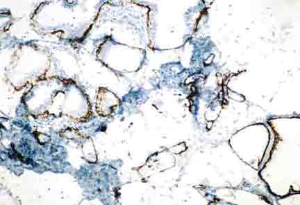
Figure 8. Low magnification of immunostaining with SMM antibody shows how the tissue has lifted off of the glass slide making the staining very unclear and difficult to interpret, original magnification 100x.

Figure 9. Medium magnification of SMM antibody strongly staining, with circumferential appearance, the myoepithelial cell layer of this non-invasive breast case, original magnification 100x.

Figure 10. Medium magnification of a strongly stained blood vessel by SMM antibody, original magnification 200x.
P63 ANTIBODY
Showed negative staining for blood vessels and background stromal myofibroblasts in all of the breast cases. P63 antibody showed very clear and clean staining and was generally easier to interpret compared with the other 4 antibodies.

Figure 11. Medium magnification of immunostaining with p63 antibody is very clear and clean although discontinuous, original magnification 200x.

Figure 12. Immunostaining of p63 (nuclear stain) identifying myoepithelial cells, note how the staining of the myoepithelial cells is very "scattered" making it more difficult to interpret the tissue section, normal magnification 200x.

Figure 13. Medium magnification of p63 antibody moderately staining epithelial cells, normal magnification 200x.
Continued...